DR V SAMPATH
HOD
I, Dr. V. Sampath, currently serve as Director and Professor of Dermatology in the Institute of DVL at Madras Medical College, Chennai, with over 29 years of extensive teaching and clinical experience in dermatology. I completed my MBBS in 1989 and MD (Dermatology) in1997 and have since held academic positions across several esteemed medical institutions, including Madras Medical College, Stanley Medical College, Kilpauk Medical College, Kanyakumari Medical College, and Chengalpattu Medical College. Over the years, I have actively contributed to medical education by teaching, mentoring students, and delivering lectures on diverse dermatological topics. My research contributions include publications on chronic spontaneous urticaria, post-biologic management of pemphigus patients, and clinicopathological studies of cutaneous tuberculosis in reputed dermatology journals. I have also completed the Revised Basic Course Workshop in Medical Education Technology, strengthening my commitment to academic excellence. With expertise in clinical dermatology, research, and education, I remain dedicated to advancing patient care and nurturing the next generation of dermatologists.
DR PUNITHAVATHI
PROFESSOR
I am currently working as Professor in the Institute of Dermatology, Venereology, and Leprosy at Madras Medical College, with over 20 years of teaching and clinical experience. I have been actively involved in undergraduate and postgraduate teaching, research activities, and patient care. I have contributed to the academic growth of the department through regular lectures, clinical training, and mentorship. My commitment to medical education and clinical excellence continues to guide my professional efforts.
DR SARADHA
PROFESSOR
I, Dr. K.P. Saradha, currently serving as Professor in the Institute of Dermatology, Venereology and Leprosy at Madras Medical College, take this opportunity to present myself-appraisal. With over three decades of academic and clinical experience, I have consistently contributed to the field through teaching, patient care, and scholarly work. I have published 4 research articles in reputed international indexed journals, demonstrating a commitment to evidence-based practice and academic advancement. In addition, I have contributed to academic literature by authoring chapters in books relevant to my specialty. I have also actively participated in academic forums, delivering oral presentations at two state-level conferences. My dedication to continued medical education is reflected in my participation in key faculty development programs like the Basic Course Workshop in Medical Education Technologies. I aim to continue contributing meaningfully to medical education, research, and clinical excellence.
DR MADHU
PROFESSOR
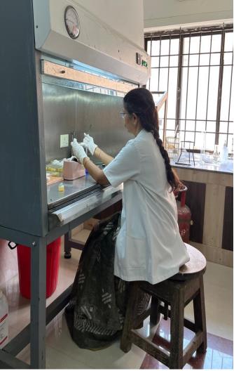
I, Dr R. Madhu am working as Professor, Institute of DVL, in charge of the Dermato mycology section , which is a referral unit for the diagnosis of subcutaneous and opportunistic mycoses, since 15 years. It has been my proud privilege to represent my Institution as a dermato- mycology faculty in the National and International conferences. I am also the nodal officer for teleconsultation, which paves way for the patients to consult doctors, sitting in the comfort of their homes. I am involved in the welfare activities of undergraduate students in the capacity of financial advisor.
DR RAJKUMAR
PROFESSOR
I, Dr. RajKumar Kannan, currently serve as Professor in the Institute of Dermatology, Venereology, and Leprosy at Madras Medical College and Rajiv Gandhi Government General Hospital, Chennai. I hold an MBBS, DDVL, MD (DVL), and IFAAD, with my undergraduate degree from Tanjore Medical College and postgraduate training at Madras Medical College. My areas of expertise include psoriasis, autoimmune bullous diseases, connective tissue disorders, and advanced cosmetology techniques such as radiofrequency, chemical peels, Botox, fillers, and lasers. Throughout my academic career, I have been recognized with several prestigious accolades ,including multiple university ranks during MBBS and gold medals for outstanding therapeutic paper presentations at national dermatology conferences. I have presented award-winning research papers on topics such as psoriasis, fungal infections, photodermatoses, and leprosy at various national and international conferences, including Dermacon and the World Congress on Pediatrics and Neonatology. My work reflects a strong commitment to research, clinical excellence, and advancing dermatological sciences.
DR MURUGAN
PROFESSOR
I, Dr. S. Murugan, am currently working as a Professor in the Institute of Dermatology, Venereology, and Leprosy at Madras Medical College. I have over 18 years of comprehensive teaching and clinical experience in reputed institutions including Madras Medical College, Chengalpattu Medical College, Madurai Medical College, and Government Villupuram Medical College. I joined Madras Medical College in March 2025 as a Professor and have consistently fulfilled my role as a full-time teacher from 9:00 AM to 2:00 PM, contributing actively to undergraduate and postgraduate teaching, clinical care, and departmental administration. I have published several indexed research papers in reputed journals including PubMed and Index Copernicus and participated in cuticon 2006 and pediatric dermatology quiz and won 2nd prize. I have presented papers in regional , state and national conferences. Chaired sessions in National and state conferences . I affirm that I am not associated with any other medical college in any capacity and have adhered strictly to the regulations laid down by the Medical Council of India and The Tamil Nadu Dr. MGR Medical University. I declare that all details provided by me are accurate and authentic to the best of my knowledge.
DR SAMEUL JAYARAJ DANIEL
PROFESSOR
I am Dr. S.J. Daniel, currently serving as Professor of Dermatology, Venereology &Leprosy (DVL) at Madras Medical College. With an MBBS from Stanley Medical College and an MD (DVL) from Madras Medical College, I have consistently committed myself to both clinical excellence and academic contribution. I have been honored with prestigious recognitions including the Doctors Day Award (2019) by the Tamil Nadu Government and the SPARSH Award for contributions to leprosy work (2021). My academic footprint includes over 30 publications in indexed journals, as well as active participation in state and national conferences through paper presentations and workshop conduct. My core areas of interest include radiosurgery, LASER surgery, vitiligo surgery, pigmentary disorders, and dermoscopy. I strive to blend patient care with innovation and knowledge dissemination, continuously enhancing my impact in the field of dermatology.
Dr.B VIJAYALAKSHMI
ASSOCIATE PROFESSOR
I am currently serving as an Associate Professor in the Institute of Dermatology at Madras Medical College, with over 17 years of dedicated teaching experience. Throughout my academic career, I have remained committed to both undergraduate and postgraduate education, fostering clinical acumen and research aptitude among students. My contributions extend beyond teaching into research, with key publications including a clinical study on vitiligo and an analysis of the cutaneous manifestations of systemic lupus erythematosus. I strive to integrate evidence-based medicine into clinical teaching and am actively involved in departmental academic activities, aiming to contribute meaningfully to the advancement of dermatological education and patient care.
DR VNS AHAMED SHARIFF
ASSOCIATE PROFESSOR
I am currently working as an Associate Professor in the Institute of Dermatology ,venereology and leprosy with a teaching career spanning over 17 years at reputed institutions including Madras Medical College. My academic journey has been enriched through consistent involvement in clinical teaching, mentoring, and research. I have contributed to several publications covering topics such as chronic spontaneous urticaria, Blaschko-linear dermatoses, renal cell carcinoma, and antimicrobial resistance patterns in N.gonorrhoeae. I remain committed to advancing dermatological knowledge through both clinical work and academic collaboration..
DR NITHYA GAYATHRI DEVI
ASSOCIATE PROFESSOR
I serve as an Associate Professor in the Institute of Dermatology, Venereology, and Leprosy at Madras Medical College, with 13 years of teaching experience. My academic interests lie-in clinical dermatology, and I have undertaken research on linear dermatoses. I have also completed the Basic Course in Biomedical Research (BCBR), strengthening my foundation in research methodology. I remain dedicated to delivering quality teaching and actively contributing to both clinical and academic growth in the department.
DR P SARASWATHY
ASSOCIATE PROFESSOR
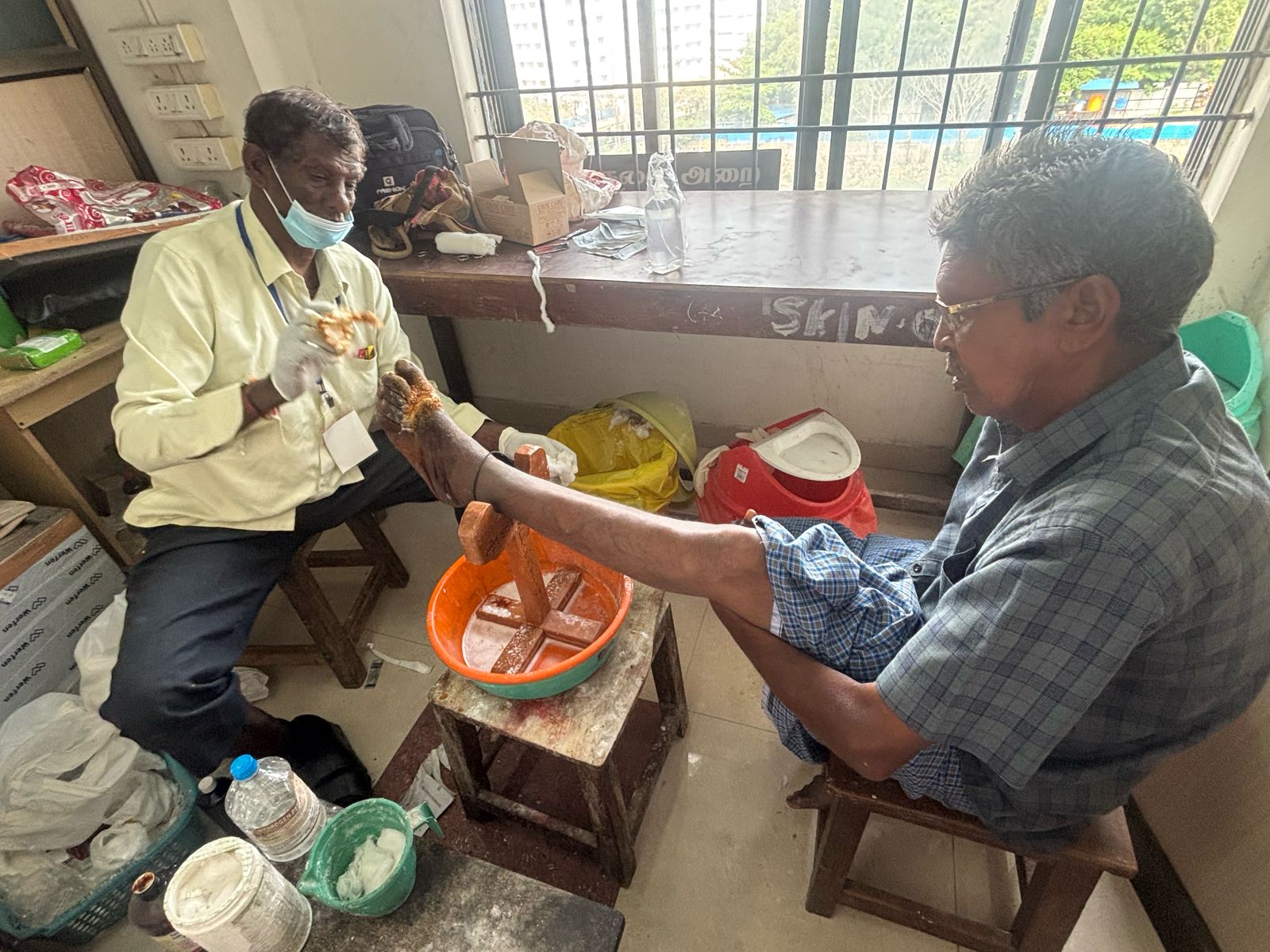
I, Dr. P. Saraswathy, am currently serving as an Associate Professor at Madras Medical College (MMC). I completed my MBBS (1995–2001), DGO (2002–2004), and MDDVL(2012–2015) from MMC, building a strong academic foundation in dermatology and related fields. I am a Central Core Member of IADVL (2024) and a Life Member of the IADVL Academy since 2004. I have served in key leadership roles, including Vice President of TNIADVL (2023) and Joint Secretary of IADVL (2022). My organizational contributions include being part of the organizing team for the GRR Quiz, cosmetology workshops at Stanley Medical College (2021–2024), and the CUTICON TN Conference (2019). My research contributions are reflected through several publications in reputed journals, covering topics such as cutaneous manifestations of lupus erythematosus, linear dermatosis, patterned wound healing using epidermal growth factor in leprosy trophic ulcers, PRP in leprosy trophic ulcers, and management of challenging pemphigus vulgaris cases with modified pulse therapy. I have also authored studies on the clinical spectrum of childhood dermatoses during peak summer, onychomadesis associated with Hand, Foot, and Mouth Disease, and the epidemiology of pediatric leprosy in a tertiary care institute.
DR K DEEPA
ASSOCIATE PROFESSOR
I am an Associate Professor in the Department of Dermatology at Madras Medical College, with over 10 years of teaching experience. I have been actively involved in research, including studies on clinical epidemiology of skin disorders, patch testing in OCD clinics, and the role of Pap smear screening in a teaching hospital. I also have a strong interest in dermatosurgery and have performed various laser procedures including aesthetic procedures as part of my clinical practice. It remain committed to integrating clinical research with quality medical education and patient care.
DR PRABAHAR P
ASSOCIATE PROFESSOR
I am an Associate Professor in the Institute of Venereology atMadras Medical College with 18 years of teaching experience. My research includes key studies on sexually transmittedinfections in MSM populations and VDRL reactivity patterns. With first-author publications in peer-reviewed journals, I strive to blend clinical expertise with academic inquiry and continue to contribute actively to teaching, research, and STI prevention strategies in a tertiary care setting.
DR DURGAVATHI C
ASSISTANT PROFESSOR
I, Dr. Durgavathi C, currently working as Assistant Professor in theInstitute of Dermatology, Venereology and Leprosy at Madras MedicalCollege, have served in this institution since June 2018, with a totalteaching experience of approximately 7 years and 11 months. Myqualifications include MBBS from IRT Perundurai Medical College (2003),MD DVL from Madras Medical College (2012), and DM DVL from thesame institution (2018), all under The Tamil Nadu Dr. M.G.R. MedicalUniversity. I have contributed actively to research and academics, withtwo research publications in indexed journals published in 2024. Additionally, I have completed key academic enhancement courses including the Basic Course in Medical Education Technology (Sri Ramachandra Medical College, 2020) and the Biomedical Research Course (ICMR/NIE, 2021). My focus remains on advancing academic standards, mentoring undergraduates and postgraduates, and continuing research in clinical dermatology.
DR S VENKATESAN
ASSISTANT PROFESSOR
I, Dr. Venkatesan S., currently serving as Assistant Professor in the Institute of Dermatology, Venereology, and Leprosy at Madras Medical College, bring over 13 years of extensive teaching experience across prestigious institutions including Stanley Medical College, Royapettah Government Hospital, and Thiruvarur Medical College. I hold an MBBS from Thanjavur Medical College (1997), followed by Diploma in Venereology(DSTD) from Madras Medical College (2002), and further specialization through DNB(DVL) from Madras Medical College(2005), all affiliated with The Tamil Nadu Dr. MGR Medical University. I have contributed significantly to academic research, with multiple publications in reputed journals, and have completed certified basic courses in Medical Education Technology and Biomedical Research. My continued academic commitment is reflected through a combination of clinical excellence, active participation in research, and dedicated mentorship to undergraduate and postgraduate medical students
DR H DHANASELVI
ASSISTANT PROFESSOR
As an Assistant Professor in the Institute of Dermatology, Venereology, and Leprosy at Madras Medical College since July 2016, I have consistently demonstrated commitment to academic excellence, patient care, and research. With over 9 years of teaching experience, including extensive senior residency across esteemed institutions, I have honed my clinical and teaching skills. I hold an MBBS from Government Medical College, Trichy and an MD from Madras Medical College, affiliated with The Tamil Nadu Dr. MGR Medical University. I have actively contributed to academic research, with four publications in peer-reviewed journals, and participated in certified basic courses on Medical Education Technology and Biomedical Research. My focus remains on advancing medical knowledge, and nurturing future medical professionals through evidence-based teaching.
DR VIDHYA C
ASSISTANT PROFESSOR
Assistant Professor, Institute of Dermatology, Venereology &Leprosy, Madras Medical College Passionate dermatologist with good clinical acumen An academically driven faculty member with 14 years of teaching and clinical experience in the field of dermatology. Proficient in Aesthetic medicine and procedural dermatology. Has contributed significantly to research with original articles in various journals related to Dermatology and venereology.. Has contributed significantly to research, with original articles on seroprevalence of HIV, HBV, HCV, and syphilis among STI patients and a clinicopathological study of cutaneous tumors of head and neck. Actively involved in STI clinics and diagnostic dermatopathology, blending academic knowledge with clinical relevance.
DR R RAJESHWARI
ASSISTANT PROFESSOR
I am Dr. Rajeswari, currently working as an Assistant Professor in the Institute of Dermatology. I have over a year of teaching and clinical experience and have actively participated in national and state-level dermatology conferences. I have presented papers at CUTICON Tamil Nadu 2020 and DERMACON 2021, focusing on clinical dermatology and case-based discussions. I am passionate about academic involvement and constantly strive to enhance my teaching and clinical skills through continued participation in professional forums.
DR S DEVASENA
SENIOR RESDIENT
I am Dr. S. Devasena, currently serving as a Senior Resident in the Department of Dermatology, Venereology & Leprosy (DVL)at Madras Medical College since September 2019. With over 18years of teaching experience in government medical institutions across Tamil Nadu, I have served in key roles including Casualty Medical Officer, Assistant Surgeon, and Tutor. I have actively contributed to academic research with multiple publications in reputed dermatology journals and have completed essential training courses . I am committed to continuous academic growth, clinical excellence, and contributing meaningfully to undergraduate and postgraduate medical education..
DR MOHAMMED MUBARAK M.S
SENIOR RESDIENT
I am Dr. Mohamed Mubarak M.S., currently working as a Senior Residentin the Department of Dermatology, Venereology & Leprosy (DVL) atMadras Medical College. I completed my MBBS from Sir SalimullahMedical College under the University of Dhaka in 2006 and subsequentlyearned my MD in Dermatology from The Tamil Nadu Dr. M.G.R. MedicalUniversity in 2015. With a total teaching experience of over 11 years and8 months, I have served as a Junior and Senior Resident across premiermedical institutions including Stanley Medical College and MadrasMedical College. I have completed foundational training through theBasic Course in Medical Education Technology and Biomedical Research. I am dedicated to providing evidence-based patient care while actively engaging in academic responsibilities and skill development to further enhance my contributions to the field of dermatology.
DR RASHMI
SENIOR RESDIENT
I am a Senior Resident in the Institute of DVL at MMC with over 7 years of teaching experience. I have special interest in teaching the students and have conducted various seminars. I have also worked with meritorious professors who expertise in dermatology and venereology and gained experience. I have also been into administrative side, helping the department and hospital in various aspects
DR S YUVARAJ
SENIOR RESDIENT
I am currently working as a Senior Resident in the Department of Dermatology, Venereology, and Leprosy at Madras Medical College. With prior experience as a Junior Resident at Govt. Stanley Medical College, I have been actively involved in clinical care, academic teaching, and departmental responsibilities. I continue to stay committed to improving patient care and medical education through evidence-based practice and active participation in academic programs..
DR PRIYA
SENIOR RESDIENT
I am currently working as a Senior Resident in the Department of Dermatology, Venereology, and Leprosy at Madras Medical College. I have actively contributed to academic and clinical services and have published one international and national journal article. These publications reflect my ongoing commitment to academic growth, evidence-based practice, and dermatological research.
DR PRAVEEN
SENIOR RESDIENT
I am Dr. Praveen M, currently serving as a Senior Resident in the Department of Dermatology, Venereology, and Leprosy at Madras Medical College, having joined in June 2024. I completed my MBBS in2009 and MD (DVL) in 2019 from Madurai Medical College, and furthered my academic growth .With over 1 year and 9 months of total teaching experience including postings at GMCH Theni and Madurai Medical College, I am actively involved in both clinical duties and academic responsibilities. I have completed certified basic courses in Medical Education Technology and Biomedical Research through MCI and ICMR respectively. My academic trajectory reflects a strong commitment to continuous learning and contribution to the dermatology department through quality patient care, teaching, and skill enhancement